I want to host a screening event
I WANT TO HOST A SCREENING EVENT >
Applying the lotus-effect to liver cancer research
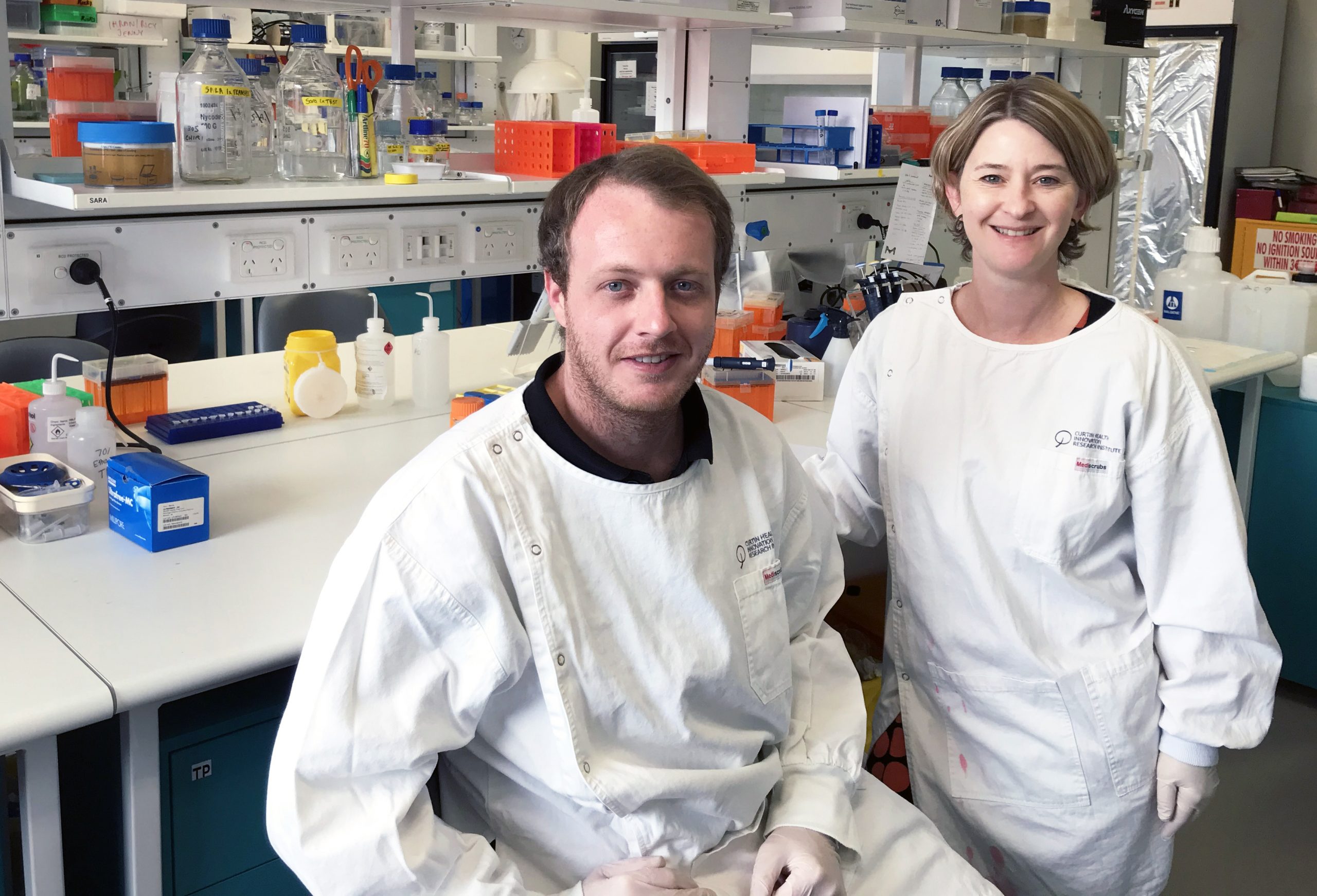
Just like the liver functions to detoxify the body, the self-cleaning abilities of the lotus flower, known as ‘the lotus-effect’, are a symbol of long life and health. The inaugural recipient of the Lions-Lotus PhD scholarship, Nathan Main, is aiming to improve both the lives and health of chronic liver disease patients through his research project at the Curtin Health Innovation Research Institute (CHIRI).
Supported by the Lions Cancer Institute (WA) Inc., Nathan’s PhD project is supervised by CHIRI’s Associate Professors Nina Tirnitz-Parker and Pieter Eichhorn, from Curtin University’s School of Pharmacy and Biomedical Sciences, and WA gastroenterologist Professor John Olynyk.
Nathan’s research is focused on decoding the language cells use to communicate, which may contribute to cancer initiation. If found to be significant to liver disease, these cells may be targeted with therapeutics to block liver cancer formation.
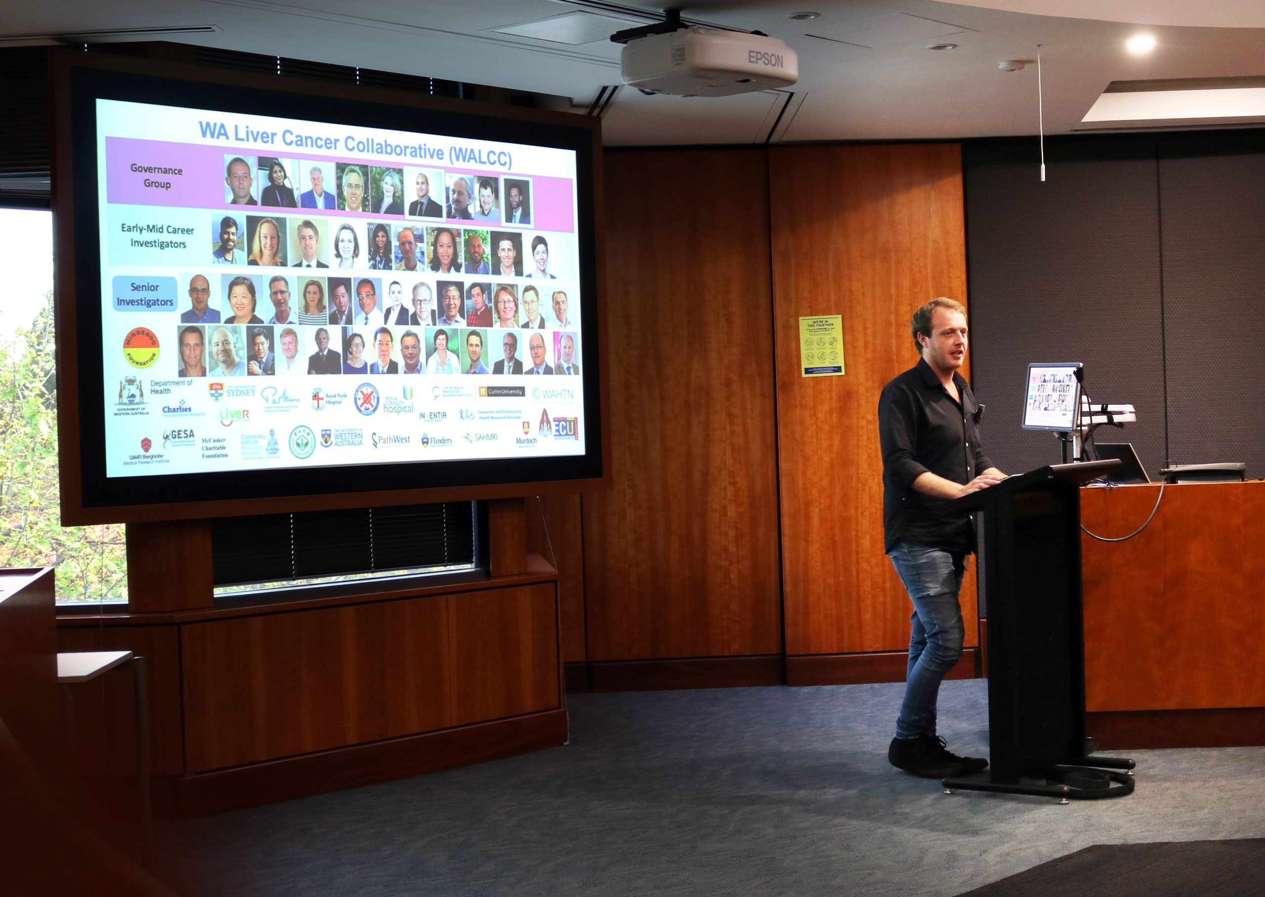
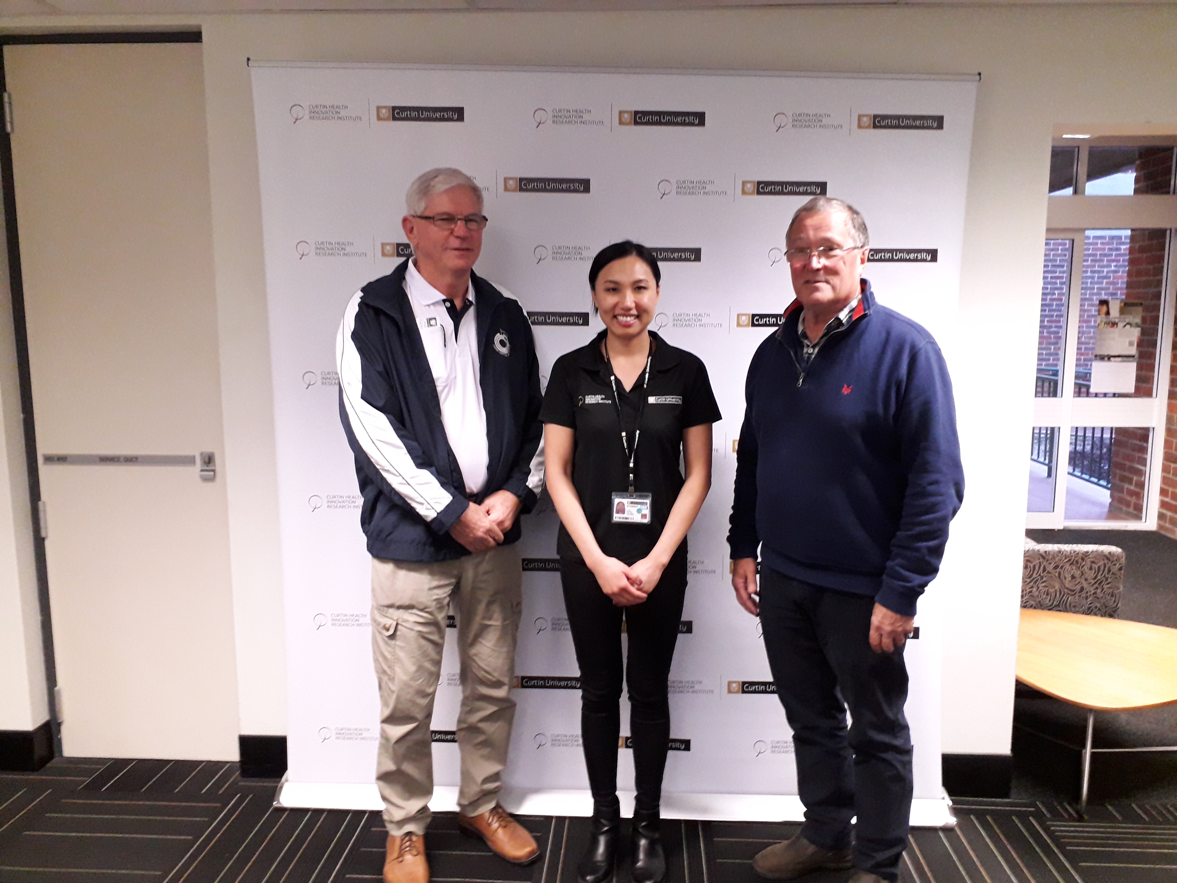
Nathan recently presented on his PhD project to representatives from the Lions Cancer Institute (WA) Inc., CHIRI and Curtin’s Faculty of Health Sciences and School of Pharmacy and Biomedical Sciences, at a special ceremony announcing his award, as the inaugural Lions-Lotus PhD Scholarship recipient.
“Ultimately, I hope my research will result in new therapeutic approaches to liver cancer, including earlier detection to improve patient outcomes, and better tolerated and more effective treatment options,” said Nathan.
Nathan Main presenting on his PhD project at a special ceremony announcing his award, as the inaugural Lions-Lotus PhD Scholarship recipient.
“I am hoping that by investigating the communication or signalling between liver cells, I will identify novel therapeutic targets to prevent the progression of chronic liver disease to liver cancer and discover biomarkers that can tell us which patients are more likely to develop a tumour.”
Nathan’s appointment to CHIRI’s Liver Disease and Regeneration Group follows his work as a Research Assistant for the Gastroenterology and Liver Laboratory at the Ingham Institute for Applied Medical Research in Sydney. Nathan is pleased to be back in Perth, having grown up and completed his undergraduate degrees there and is looking forward to applying his knowledge and expertise in the field of liver disease research to his PhD project.
CHIRI Director Professor John Mamo said, “Nathan’s highly-anticipated appointment to CHIRI will help Nina and her research group further explore the potential of their recent research findings, aimed at improving outcomes for liver disease and cancer patients. We can’t thank the Lions Cancer Institute (WA) Inc. and Nina enough for pioneering this scholarship program and supporting student and research excellence at our institute. Nathan’s research will contribute to important and ongoing research by Nina and her Liver Disease and Regeneration Group at Curtin-CHIRI, aimed at finding new treatments for liver disease and improving the lives of patients and their families.”
Liver Disease and Regeneration Group researchers recently identified a ‘goldmine’ of genetic markers and protein receptor-ligand pairs that are possible contributors and predictors of liver cancer and provide prime candidates for Nathan’s research. Nina said with these findings, along with the recent announcement of a $10.8 million boost for primary liver cancer research in WA, Nathan’s appointment could not be more timely.
“Nathan’s appointment comes at a time of unprecedented support for primary liver cancer research in our state, making this a great opportunity to progress both his career and our research,” Nina said.
Nina and Professor Peter Leedman from the Perkins Institute and UWA will co-lead the WA Liver Cancer Collaborative team at a new $10.8 million primary liver cancer research centre for Perth (read the announcement here).
“The establishment of this state-wide collaborative program will provide Nathan with access to a large, multidisciplinary network of clinicians and scientists to guide his research, as well as access to world-class facilities, technologies and clinical samples,” Nina said.
“It’s an exciting time for our research team and for Nathan and I am grateful to the Lions Cancer Institute (WA) Inc. and CHIRI for this opportunity to nurture the next generation of research excellence through a scholarship dedicated to researching liver disease and cancer.”
You can find out more about the Lions Lotus PhD scholarship here, or give to the program here.
Postgraduate Research Scholarships
“The Lions Cancer Institute (WA) Inc. continues to support PhD Students involved with all types of cancer research. We can only wish these dedicated young people every success and hope that our continued financial support will one day lead to a better understanding and possible cure?”
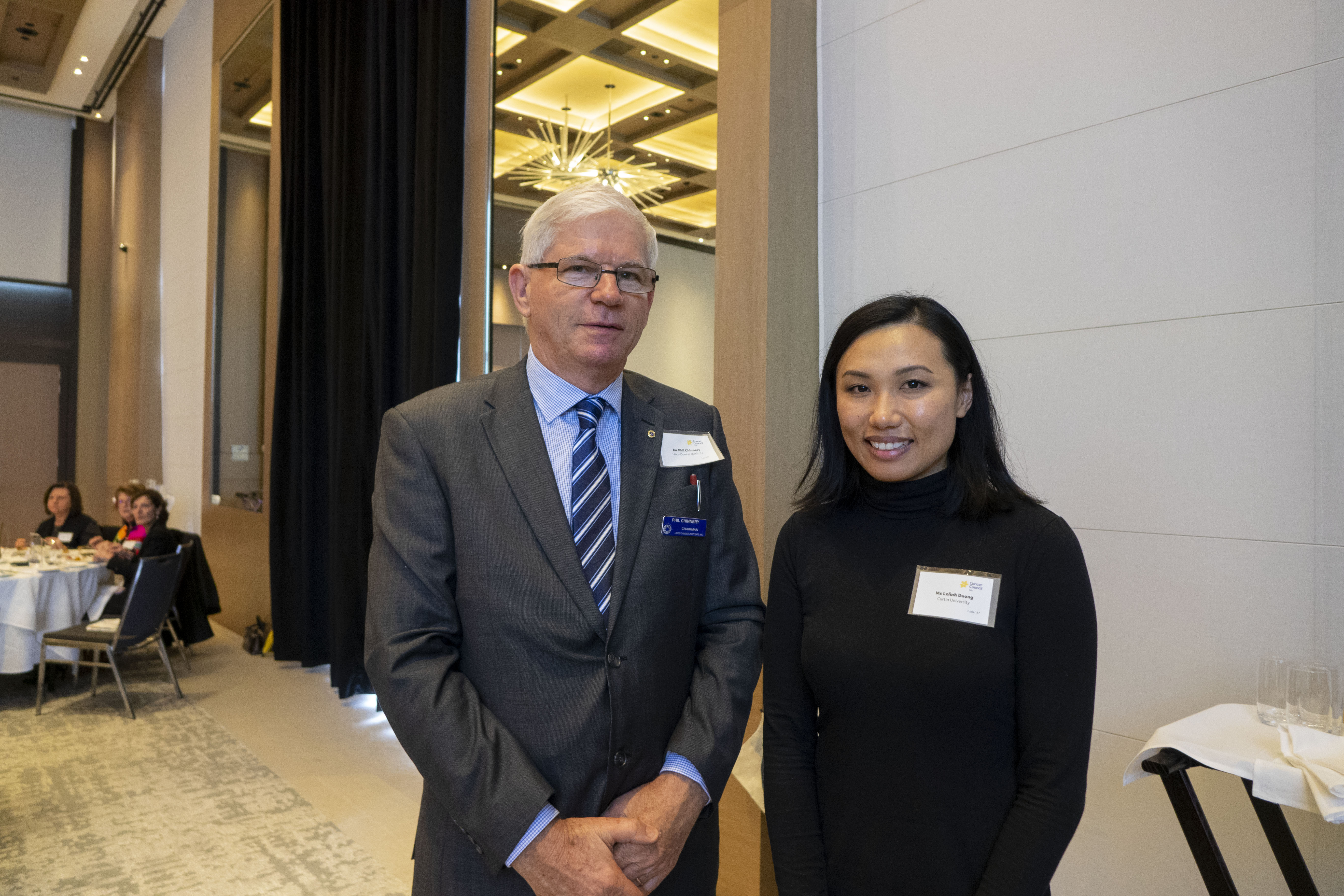
— Phil Chinnery, LCI Chairman
The Lions Cancer Institute (WA) Inc. has for many years supported by way of a Postgraduate Scholarship, “top up” financial support for a selected student studying with the University of Western Australia to undertake further study over three years to attain either a Honours or PhD degree, specializing in the study of cancer causes and towards a cure.
The Scholarship, instigated and supported by the Lions Club of Armadale Kelmscott and assisted by other Lions Clubs in 1995, was set up in memory of the late wife and son of current Chairman, Lion Phil Chinnery. Karen and Joshua both died from a brain tumour in 1992 and 1995 respectively.
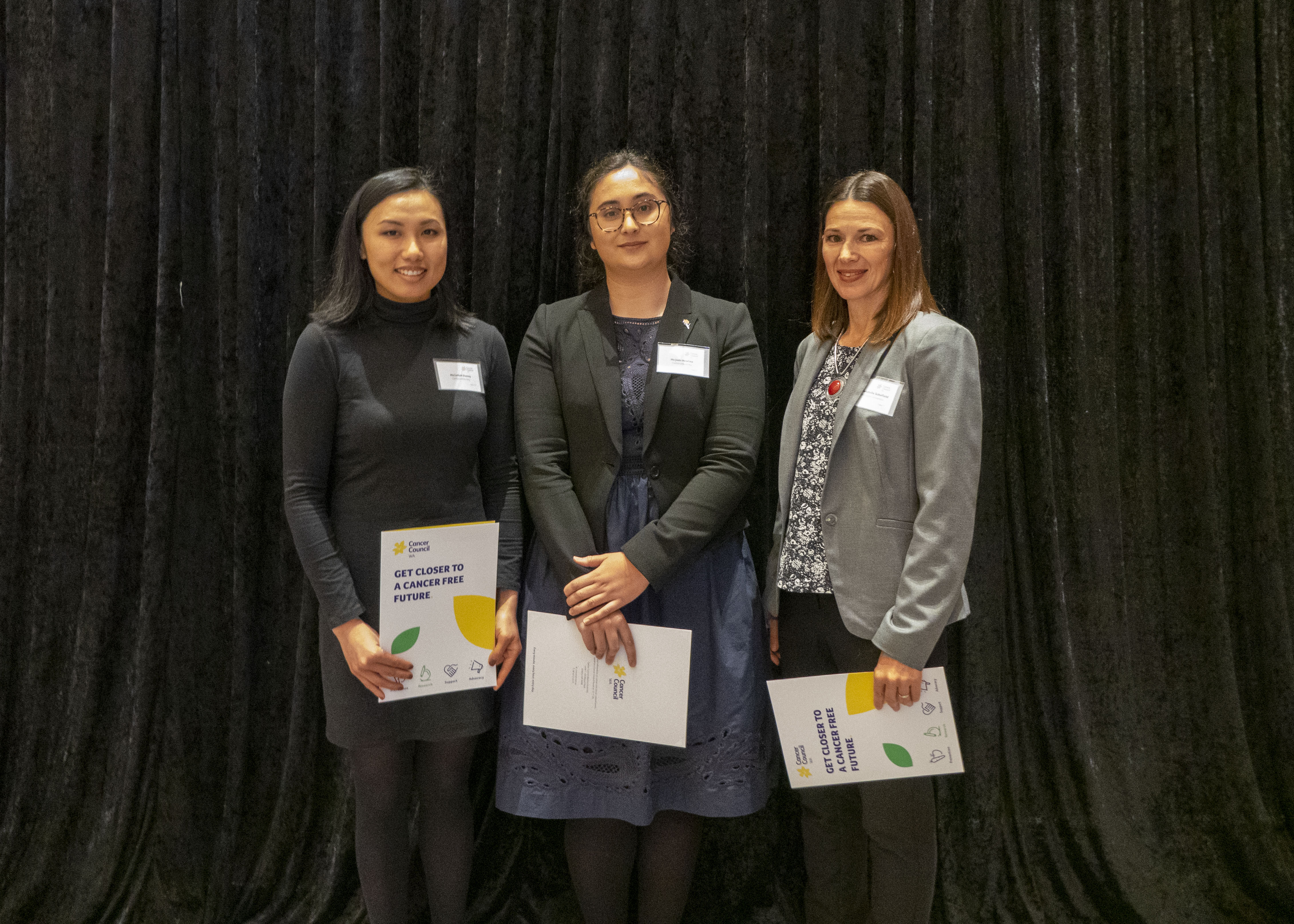
The Lions Cancer Institute (WA) Inc. is determined to continue the support of students such as Tracey, Jade and Lelinh in the search for better treatments and hopefully a cure for cancer. Up to now only one student has been able to be supported over the three-year course of study. We are very happy to have been able to increase this to two students studying concurrently. Of course, this costs money and we appeal to all to consider donating to the scholarship to help keep this essential study going.
Tracey Seymour is the ninth student assisted by the Lions Cancer Institute (WA) Inc. over the past 22 years through the Karen and Joshua Chinnery Memorial Scholarship, managed by the Institute. Tracey will complete her studies in early 2019.
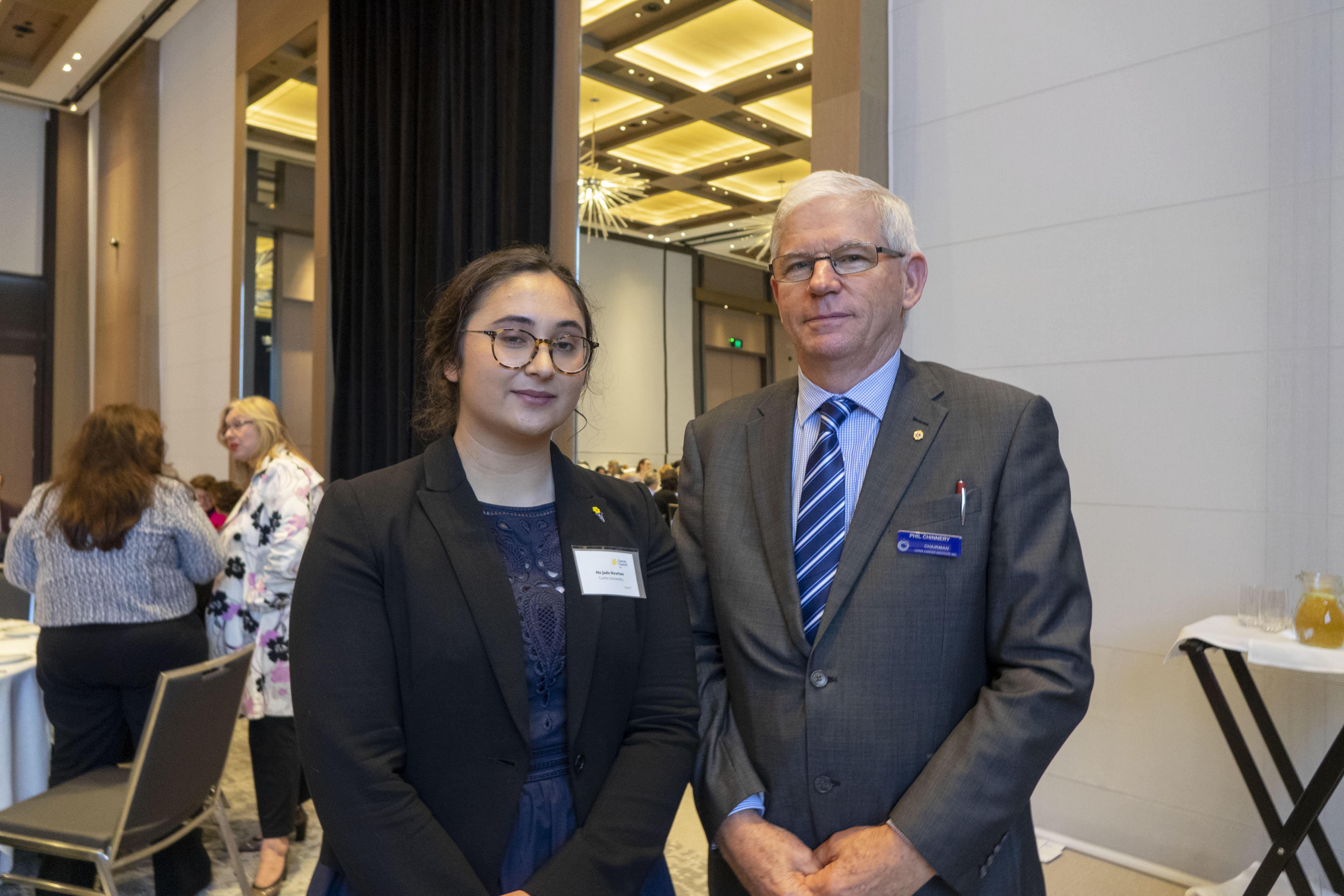
Two new students have been selected from recommendations made by Cancer Council WA to commence in 2019, being Ms. Jade Newton (3-year term) and Ms. Lelinh Duong (1-year term). Both Jade and Lelinh are students of Curtin University.
PhD Students Cancers Research's Reports
Lelinh Duong, 2nd Report, 2020
PhD student from The Curtin University
Investigating how key immune cells, known as macrophages, contribute to tumour growth in the elderly and impair response to therapy
Background and Aims:
Cancer incidence and severity increases with age. It is estimated that people aged 60 years and older account for 86 percent of cancer-related deaths (Australian Institute of Health and Welfare, 2019). However, the elderly population are often overlooked in pre-clinical studies and cancer treatment options are limited and may not be effective. Therefore, there is a crucial requirement for better therapeutic options for elderly cancer patients. Many studies suggest that the impairment of the immune system with ageing may contribute to poorer anti-cancer outcomes in elderly people. Macrophages are an immune cell type which can help fight cancer cells. However, macrophages can be easily persuaded to help tumours grow. Many tumours can be made of up to 50 percent macrophages and the number is associated with poor clinical outcome. Currently, very few studies have looked at the effect of ageing on macrophages in the tumour.
My project aims to 1) investigate the mechanisms that cause a key immune cell, called a macrophage, to become more tumour-promoting with age. 2) These components will be targeted with therapeutic drugs in hopes to restore immune balance and improve anti-cancer responses in the elderly.
Summary of Findings
Aim 1
Using a mouse model of mesothelioma and lung cancer, my research has identified changes to communication molecules inside macrophages found in the tumour, that are instrumental in ageing and cancer. These molecules can guide the function of macrophages, such as promote tumour growth and contribute to inflammation in the tumour. We found that as tumours progressed, the activity of these communication molecules increased. This occurred faster in the elderly, which may explain why we see faster tumour growth in the elderly compared to young hosts. Importantly, these molecules are promising targets to potentially restore immune balance and improve anti-cancer outcomes in the elderly.
The findings from aim 1 were presented at several local and national conferences and won the following awards:
- Overall Poster Award at the 48th Annual Scientific Meeting of the Australian and New Zealand Society for Immunology, Adelaide; 12th December 2019
- BD Science Communication Award at the 48th Annual Scientific Meeting of the Australian and New Zealand Society for Immunology, Adelaide; 12th December 2019
- Campion Ma Playoust Memorial Award at the Australian Society for Medical Research 58th National Scientific Meeting, Fremantle; 21st November 2019
- Prize for best oral presentation at Mark Liveris Seminar, Curtin University; 27th September, 2018
- Best Student Oral Presentation at the WA Combined Biological Sciences Meeting, University of Western Australia; 31st August, 2018
Aim 2
In recent experiments, we blocked the communication molecules identified in Aim 1 using drugs already approved for clinical use. Our initial findings show this to be effective in slowing tumour growth in the elderly. Next, we will validate the findings and expand this work to confirm the drugs are indeed targeting the impaired macrophages and inhibiting their ability to promote tumour growth.
Significance
So far, the findings from this research have advanced our knowledge of cancer progression by identifying some of the mechanisms involved. Furthermore, we have identified promising therapeutic targets that can potentially improve anti-cancer outcomes for elderly people. This project aims to help guide future cancer research to ultimately improve anti-cancer outcomes, such as slow tumour growth, improve response to therapies, and increase survival for people with cancer.
Jade Newton, 2nd Report, 2020
PhD student from The Curtin University
Project title
Are Western Australian advanced cancer patients’ psychosocial support needs being met? A mixed methods investigation of the provision and utilisation of psychosocial support for advanced cancer patients with non-haematological malignancies.
Summary
Some people are diagnosed with cancer that has spread to other parts of their body and they are not able to be cured. This is ‘advanced cancer’. We don’t know a lot about how people with advanced cancer manage the emotional and social (or, ‘psychosocial’) impact of their diagnosis, and how available support affects their experience.
This project will explore what psychosocial support people with advanced cancer use, how it impacts them, and the costs and benefits of using psychosocial supportive care. This will be achieved by looking at what research has been done in this area [study one], interviewing people with advanced cancer to learn about their experiences [study two], and undertaking a cohort study, where we will survey patients over several months to look at their needs, distress, and support services used over time [study three].
Understanding the needs and service use of people with advanced cancer will help to make sure we are providing the best support to lessen the distress and suffering they may experience.
Project update – July 2020
Study one is underway. I have finished searching the literature, and am currently reviewing guidelines and previous research that has explored the how support is provided to cancer patients.
Data collection has been completed for study two. Twenty-three people living with advanced cancer kindly shared their experiences living with their diagnosis, and discussed the kinds of support they have needed and used. Interviews were completed at the participants’ convenience, at home, over the phone, or at hospital during treatment. We found that in Western Australia, whilst there a lot of resources available to support people, they were not always available for people with advanced cancer. Some participants experienced barriers to support including suitability of support (e.g. face-to-face support vs. phone based), distance needed to travel to get to support services, availability of health professionals such as social workers, and ability to access financial support. A short report will be prepared to share these findings with local support services, so that they are able to address these barriers to support. This research has been accepted for a poster presentation at the upcoming NVivo Virtual Conference, which will be online due to the pandemic. A paper describing participant experiences accessing support services is also being finalised for submission to an academic journal.
Study three is still in the planning stages and we need to apply for ethics approval prior to commencing the study.
Tracey Seymour, 4th Report, 2019
PhD student from The University of Western Australian and Telethon Kids Institute
My PhD research focuses on finding more effective therapies for the most lethal human brain tumour, glioblastoma. Current treatment for glioblastoma includes safe maximal surgical resection, followed by radiotherapy and chemotherapy such as temozolomide. However, these treatments are not curative and patients only rate of survival is between 12 to 15 months from diagnosis. A reason for these treatments not being curative is that glioblastoma cells having the ability to resist the damaging effects of radiotherapy and temozolomide.
My PhD research involves pre-clinical studies that is currently testing a therapy, in hope that it will improve patient outcomes and extend survival.
Current research aims:
1. Determine whether new treatments will interact together with radiotherapy and/or chemotherapies such as temozolomide and gemcitabine.
2. Explore the mechanism in which these new therapies help to improve tumour cell death.
3. Examine whether combining new therapies to radiation therapy or chemotherapies such as temozolomide and gemcitabine will provide a survival benefit.
Aim 1
Determine whether new treatments will interact together with radiation therapy and/or chemotherapies such as temozolomide and gemcitabine.
Background
The standard of care for glioblastoma includes safe maximal resection, followed by radiotherapy and chemotherapy with temozolomide. This treatment protocol is not curative and tumour growth is still very rapid with a mean survival rate of only 12 to 15 months. Furthermore, glioblastoma has shown resistance to radiotherapy and temozolomide through the ability of repairing the DNA damage induced by these therapies. This particular study aims to prevent the DNA damage repair pathway with new therapies to enhance glioblastoma cell death.
Findings
Using several human glioblastoma cell lines, we have tested three new drug therapies. These therapies were tested alone or in combination with radiotherapy, or chemotherapies temozolomide and gemcitabine. The most promising therapy was LY2606368, an inhibitor of a protein called cell cycle checkpoint kinase 1/2. We have shown that LY2606368 causes a reduction in glioblastoma cell growth. More importantly, when combined with radiotherapy or temozolomide or gemcitabine, LY2606368 was compatible, where combination therapy produced enhanced glioblastoma cell death.
Significance
This study shows that combining LY2606368 to the current treatment of glioblastoma can enhance glioblastoma cell death.
Aim 2
Explore the mechanism in which these new therapies help to improve tumour cell death.
Findings
Protein analysis, confirmed that LY2606368 stopped the activation of the protein called cell cycle checkpoint kinase 1/2. We also determined that this prevented glioblastoma cells from repairing damage caused by radiotherapy or chemotherapy such as temozolomide or gemcitabine, therefore, glioblastoma cells remain damaged and accumulated more damage. Through cell analysis we were able to determine that these damaged glioblastoma cells over time start to undergo the process of cell death.
Significance
These experiments help us understand how the LY2606368 is working to enhance glioblastoma cell death.
Aim 3
Examine whether combining new therapies to radiation therapy or chemotherapies such as temozolomide and gemcitabine will provide a survival benefit.
Current Findings
Using three different glioblastoma cell lines, we implanted these cells into the brains of mice. The combination treatment of LY2606368 with radiotherapy, temozolomide or gemcitabine reduced tumour cell growth and increased tumour cell DNA damage. Using our mice models of glioblastoma we have also shown that combining LY2606368 to radiotherapy, temozolomide or gemcitabine significantly extend survival when compared to single therapy.
Significance
LY2606368 is also known as prexasertib and is currently in clinical trial for other solid tumours such as ovarian and breast cancers. The data collected from this project is helping to establish a proposal for a future clinical trial for glioblastoma patients.
-
- Seymour T, Kuchibhotla M, Nowak A, Gottardo N & Endersby R. Improving conventional therapy for glioblastoma via cell cycle checkpoint inhibition. Lorne Cancer Conference, Victoria. February 2019. Flask Talk and Poster Presentation
- Seymour T, Kuchibhotla M, Nowak A, Gottardo N & Endersby R. Exploring cell cycle checkpoint inhibition and gemcitabine treatment for glioblastoma, Trials Group for Neuro-Oncology Scientific Meeting, Brisbane, October 2018. Oral Presentation
- Seymour T, Kuchibhotla M, Nowak A, Gottardo N & Endersby R. Exploring cell cycle checkpoint inhibition and gemcitabine treatment for glioblastoma, Combined Biological Sciences Meeting, Perth August 2018. Poster Presentation
- Seymour T, Kuchibhotla M, Nowak A, Gottardo N & Endersby R. Exploring cell cycle checkpoint inhibition and gemcitabine treatment for glioblastoma, Australian Society for Medical Research, Perth June 2018. Oral Presentation
- Seymour T, Kuchibhotla M, Nowak A, Gottardo N & Endersby R. Cell cycle checkpoint inhibition enhances conventional glioblastoma treatments. Lorne Cancer Conference, Victoria. February 2018. Poster Presentation
Lelinh Duong, 1st Report, 2019
PhD student from The Curtin University
Investigating how key immune cells, known as macrophages, contribute to tumour growth in the elderly and impair response to therapy
Background
The Australian population is ageing, and cancer rates increase with old age, including mesothelioma and lung cancer. Prognosis for these types of cancers remain poor and anti-cancer treatment options are limited and may not be effective. This may be due to the impairment of the immune system with ageing. Macrophages are an immune cell type which can help fight cancer cells. However, macrophages can be easily persuaded to help tumours grow. Many tumours can be made of up to 50 percent macrophages and the number is associated with poor clinical outcome. Currently, few studies have looked at the effect of ageing on macrophages and no studies have looked at it in mesothelioma and lung cancer. The purpose of my project is to investigate if there are changes during ageing that cause macrophages to be more tumour promoting.
Research Aims
1. To determine if macrophages become more tumour-promoting in older age
2. To assess the mechanisms by which macrophages in the tumour from the elderly
are resistant to anti-cancer therapy
Summary of Findings
Aim 1
We have found that in our mouse model of mesothelioma and lung cancer the tumours grow faster in elderly compared to young. Notably, there were significantly more macrophages in the tumours of the elderly. As tumours developed, the tumour-promoting “bad” type of macrophages increased in number and this happens quicker in the elderly compared to young.
I investigated how macrophages used sugar for energy, as this is important for their function. We found that the “bad” macrophages were consuming more sugar. Concurrently, they produced higher amounts of the molecules known to promote tumour growth, contribute to further inflammation in the tumour and instruct other macrophages to infiltrate the tumour.
Next, I looked at a communication pathway inside the cells, called mammalian target for rapamycin; which is involved in regulating energy metabolism and closely linked to how macrophages function. I found there was a breakdown in this communication pathway in macrophages in the tumour as they converted to the “bad” type. Specifically, the “brake” which prevents this pathway being overused had been lifted. This likely facilitates the excessive uptake of sugar; which may provide macrophages with the energy required to produce more molecules that can promote tumour growth. Importantly, this “brake” appears to be lifted earlier in the elderly compared to young. This could explain why we see more of the “bad” macrophages earlier during tumour progression in the elderly.
Studies are ongoing to further characterise how macrophages become more tumour-promoting in the elderly. The findings from Aim 1 will identify components that can be blocked with drugs to reverse or stop macrophages from converting to the “bad” type.
Aim 2
Our group had previously shown that macrophages in the elderly can sabotage the anti-cancer effects of immunotherapy. Aim 2 will examine how macrophages from the elderly are hindering immunotherapy as per Aim 1. I will then treat mice with immunotherapy with/without the drug as per Aim 1. These findings will identify ways we can improve immunotherapy in the elderly.
Significance
This project will help advance our understanding of how macrophages promote the development of tumours and impede on therapy effectiveness; and how it changes with age. This will help guide the design of effective anti-cancer therapies for the elderly. Additionally, these findings may be relevant to other cancers, such as prostate and colorectal. Ultimately, I hope to identify ways to improve outcomes for all cancer patients.
Jade Newton, 1st Report, 2019
PhD student from The Curtin University
Project title
Are Western Australian advanced cancer patients’ psychosocial support needs being met? A mixed methods investigation of the provision and utilisation of psychosocial support for advanced cancer patients with non-haematological malignancies.
Summary
Some people are diagnosed with cancer that has spread to other parts of their body and they are not able to be cured. This is ‘advanced cancer’. We don’t know a lot about how people with advanced cancer manage the emotional and social (or, ‘psychosocial’) impact of their diagnosis, and how available support affects their experience.
This project will explore what psychosocial support people with advanced cancer use, how it impacts them, and the costs and benefits of using psychosocial supportive care. This will be achieved by looking at what research has been done in this area, interviewing people with advanced cancer to learn about their experiences, and undertaking a cohort study, where we will survey patients over six months to look at their needs, distress, and support services used over time.
Understanding the needs and service use of people with advanced cancer will help to make sure we are providing the best support to lessen the distress and suffering they may experience.
Project update
At the beginning of 2019, I was fortunately awarded a Lion’s Cancer Institute PhD Top Up Scholarship, which has helped me to swap from studying part time whilst working three days a week to studying full time and focussing on my research.
During March to July 2019, I undertook a Health Economics unit at Curtin University, and in April 2019, I successfully applied for and received an expenses-paid trip to Melbourne to attend a workshop on using existing datasets in Australia. The skills and knowledge learned are directly applicable to the planned cohort study.
With the assistance of the Consumer and Community Health Research Network, in April 2019 we recruited two consumers to be involved with the project through providing feedback on study design and participant documents.
In May 2019, this project received faculty approval from Curtin University to start undertaking the research.
Currently, several applications are in progress to gain approval to start interviewing people about their experiences. People receiving care for an advanced cancer diagnosis will be recruited from Sir Charles Gairdner Hospital, and Saint John of God Hospital in Midland. I am also developing the protocol for a literature review which will investigate what is currently known about how organisations provide supportive care services.
I would like to thank the Lion’s Cancer Institute for their generosity and kindness, and greatly appreciate their support to undertake this work.
■ Jade Newton—PhD student from The Curtin University
Tracey Seymour, 5th Report, 2020
PhD student from The University of Western Australian and Telethon Kids Institute
Overview of my PhD project
My PhD research focuses on testing a new drug therapy, called prexasertib (LY2606368) for treatment of the most lethal human brain tumour, glioblastoma. The current treatments for glioblastoma include surgery, radiation therapy and a chemotherapy called temozolomide. These treatments are not curative and patient survival is still very poor with a median survival of 12 to 15 months from diagnosis. A reason for these treatments not being curative is that glioblastoma cells having the ability to withstand the effects of radiation therapy and temozolomide.
My PhD research involves testing prexasertib in combination with radiation therapy or temozolomide, or another chemotherapy called gemcitabine, in hope that it will improve patient outcomes and prolong survival.
Research aims:
1. Determine whether prexasertib will interact together with the current treatments of glioblastoma.
2. Explore the mechanism in which prexasertib helps to improve tumour cell death.
3. Examine whether combining prexasertib to radiation therapy or chemotherapies such as temozolomide and gemcitabine will prolong survival.
Aim 1
Determine whether prexasertib will interact together with the current treatments of glioblastoma.
Findings
We have shown that prexasertib causes a reduction in glioblastoma cell growth. More importantly, prexasertib worked together with radiation therapy or temozolomide or gemcitabine and that combination therapy further reduced glioblastoma cell growth.
Significance
This study shows that prexasertib works well with existing glioblastoma therapies and helps to kill more glioblastoma cells.
Aim 2
Explore the mechanism in which prexasertib helps to improve tumour cell death.
Findings
Protein analysis, confirmed that prexasertib stopped the activation of the protein called cell cycle checkpoint kinase 1/2. This protein is overactive in cancers and aids in protecting cancer cells from the effects of radiation therapy and chemotherapy. We also demonstrated that prexasertib in combination with radiation therapy, temozolomide or gemcitabine increased the number of dying glioblastoma cells by causing unfixable damage to DNA.
Significance
These experiments help us understand how the prexasertib works to enhance glioblastoma cell death.
Aim 3
Examine whether combining prexasertib to radiation therapy or chemotherapies such as temozolomide and gemcitabine will prolong survival.
Findings
Using three different glioblastoma cell lines, we implanted these cells into the brains of mice. The combination treatment of prexasertib with radiation therapy, temozolomide or gemcitabine reduced tumour cell growth and increased tumour cell DNA damage. Using our mice models of glioblastoma, we have also shown that combining prexasertib to radiation therapy, temozolomide or gemcitabine significantly extend survival when compared to single therapy.
Significance
Prexasertib is currently in clinical trial for other solid tumours such as ovarian, breast and most importantly paediatric brain tumours. The data collected from this project has helped to establish a clinical trial proposal that was presented to The Cooperative Trials Group for Neuro-Oncology (COGNO).
Where am I now
• I am currently writing up my thesis with a planned submission for November 2020.
• I have also started looking for employment in research in Perth.
• My PhD supervisors and I will also be writing up all this data and submitting a published paper in the Special “Glioblastoma” Issue with MDPI (Multidisciplinary Digital Publishing Institute). We expect the paper to be published in mid-2021.
Tracey Seymour, 3rd Report, 2018
PhD student from The University of Western Australia and Telethon Kids Institute
My PhD research focuses on finding new therapies for the most common brain tumour, glioblastoma. Glioblastoma is a fatal type of human brain tumour. Current treatment includes maximal safe surgical resection, followed by radiotherapy and chemotherapy such as temozolomide. But these treatments are not curative and an average survival of 15 months is very dismal. Poor patient outcomes are mainly due to glioblastoma cells having the ability to resist radiotherapy and temozolomide.
My PhD research involves pre-clinical studies that is currently testing new therapies, in hope to find therapies that will augment current treatments and improve patient outcomes.
Research aims
- Determine whether new treatments will interact together with radiotherapy and/or chemotherapies such as temozolomide and gemcitabine.
- Explore the mechanism in which these new therapies help to improve tumour cell death.
- Examine whether combining new therapies to radiation therapy or chemotherapies such as temozolomide and gemcitabine will provide a survival benefit.
Aim 1: Determine whether new treatments will interact together with radiation therapy and/or chemotherapies such as temozolomide and gemcitabine.
Current Findings
We have tested three new drug therapies. The most promising therapy tested was an inhibitor of a protein called cell cycle checkpoint kinase 1/2 (inhibitor of Chk1/2). Using several different glioblastoma cell lines, we have shown that the inhibitor of Chk1/2 causes a reduction in tumour cell growth. More importantly, this Chk1/2 inhibitor works together with radiotherapy and chemotherapies such as temozolomide and gemcitabine.
Significance
This study has helped us determine a potential new therapy for patients with glioblastoma.
Aim 2: Explore the mechanism in which these new therapies help to improve tumour cell death.
Current Findings
Using protein analysis, we confirmed that the Chk1/2 inhibitor was stopping the protein called cell cycle checkpoint kinase 1/2. We also determined that this prevented tumour cells from repairing damage caused by radiotherapy or chemotherapy such as temozolomide or gemcitabine. Therefore, tumour cells remain damaged and further studies will determine if these damaged tumour cells are undergoing the process of dying.
Significance
These experiments help us understand how the Chk1/2 inhibitor is working to improve tumour cell death.
Aim 3: Examine whether combining new therapies to radiation therapy or chemotherapies such as temozolomide and gemcitabine will provide a survival benefit.
Current Findings
Using three of our glioblastoma cell lines, we have implanted these cells into the brains of mice. Firstly, the immediate effects of the inhibitor of Chk1/2 with/without radiotherapy, temozolomide or gemcitabine was examined. Combination therapy reduced tumour cell growth and increased tumour cell DNA damage. Excitedly, in two out of three cell lines, the combination of Chk1/2 inhibitor with radiotherapy, temozolomide or gemcitabine significantly extended survival.
Significance
These studies show the potential of combining the Chk1/2 inhibitor with radiotherapy or chemotherapy in improving patient outcomes.
All of these experiments will provide robust data that will inform future clinical trials for glioblastoma patients.
Conference attendance
- Seymour T, Kuchibhotla M, Nowak A, Gottardo N & Endersby R. Exploring cell cycle checkpoint inhibition and gemcitabine treatment for glioblastoma, Australian Society for Medical Research, Perth June 2018. Oral Presentation
- Seymour T, Kuchibhotla M, Nowak A, Gottardo N & Endersby R. Cell cycle checkpoint inhibition enhances conventional glioblastoma treatments. Lorne Cancer Conference, Victoria. February 2018. Poster Presentation
- Seymour T, Kuchibhotla M, Nowak A, Gottardo N & Endersby R. Cell cycle checkpoint inhibition enhances conventional glioblastoma treatments. Telethon Kids Institute Scientific Retreat, Perth November 2017. Oral presentation.
- Seymour T, Kuchibhotla M, Nowak A, Gottardo N & Endersby R. Cell cycle checkpoint inhibition enhances conventional glioblastoma treatments. Child and Adolescent Health Services Symposium, Perth October 2017. Poster presentation.
- Seymour T, Nowak A, Gottardo N & Endersby R. Sensitising glioblastoma to enhance cancer therapy, Combined Biological Sciences Meeting, Perth August 2017. Oral Presentation
- Seymour T, Nowak A, Gottardo N & Endersby R. Sensitising glioblastoma to enhance cancer therapy, Australian Society for Medical Research, Perth June 2017. Oral Presentation
Tracey Seymour, 2nd Report, 2017
PhD student from The University of Western Australia
Currently I am researching the most common human brain tumour glioblastoma at the Telethon Kids Institute within the Brain Tumour Research Group. Glioblastoma is the most common and aggressive type of human brain tumour. Current treatment includes maximal safe surgical resection, followed by radiotherapy and chemotherapy. Despite intensive treatment, this tumour remains incurable with patients surviving only 15 months from diagnosis. Radiotherapy and chemotherapy fail to efficiently kill these tumour cells and this is partly due to the ability to repair treatment-induced damage.
The research involved in my PhD includes pre-clinical studies that focus on testing new drugs to improve the clinical outcomes and survival of those diagnosed with glioblastoma.
Current research aims
- Determine whether novel therapies will interact synergistically with radiation therapy and/or chemotherapies such as temozolomide and gemcitabine.
- Examine the survival benefits of novel therapies in addition to radiation therapy and/or chemotherapies such as temozolomide and gemcitabine using animal models.
- Determine the mechanism in which these novel therapies help to enhance tumour cell death.
Aim 1: Determine whether novel therapies will interact synergistically with radiation therapy and/or chemotherapies such as temozolomide and gemcitabine.
Current Findings
We have tested three new drug therapies; they are inhibitors of the DNA damage repair pathway and inhibit kinases ATR, Chk1/2 and Wee1. Using several different glioblastoma cell lines, we have shown that these new drugs would cause a reduction in tumour cell growth. Additionally, we have also combined these new drugs with radiation therapy, temozolomide or gemcitabine. From these combination treatments, we have shown that the inhibitor of kinase Chk1/2 interacts synergistically with radiation therapy, temozolomide and gemcitabine.
Significance
This study has helped us determine which treatment combinations will act synergistically (act together), antagonistically (act against each other) and additively (work separately). For these treatment combinations, we want synergistic interaction and not antagonistic and additive interaction as these predict potential toxicity issues. Finding synergistic interactions will set the basis for which treatment combinations will be testing in our animal models (see Aim 2).
Aim 2: Examine the survival benefits of novel therapies in addition to radiation therapy and/or chemotherapies such as temozolomide and gemcitabine using animal models.
Current Findings
Using one of our glioblastoma cell lines, we implanted these cells into the brains of mice. Once tumour was established, we examined the acute effects of radiation, temozolomide or gemcitabine with/without the inhibitor of Chk1/2. The addition of inhibitor of Chk1/2 to radiation therapy significantly reduced tumour cell growth. Furthermore, the combination of inhibitor of Chk1/2 and gemcitabine significantly increased tumour cell damage and concomitantly reduced growth. Animal studies are still ongoing, and current experiments are establishing the survival benefit of the inhibitor of Chk1/2.
Significance
These studies will establish whether the addition of these new drugs to current treatments of glioblastoma will enhance tumour cell death and most importantly improve survival. The data collected from these studies will help to inform future clinical trials with patients.
Aim 3: Determine the mechanism in which these novel therapies help to enhance tumour cell death.
Current Findings
Current studies are still ongoing and are in early optimisation stages.
Significance
These experiments will help us understand how these new drug treatments are working to enhance glioblastoma tumour cells.
Conference attendance
- Cooperative Trials Group for Neuro-Oncology (COGNO), Sydney, September 2016. Poster presentation. Seymour T, Jackson M, Tat-Lai C, Nowak A, Kakulas F. Gene expression profiling reveals stem cell signatures and therapeutic targets for glioblastoma and gliosarcoma.
- Telethon Kids Scientific Retreat, Perth, November 2016. Oral presentation. Seymour T, Nowak A, Gottardo N, Endersby E. Sensitising glioblastoma to enhance cancer therapy.
- Australian Society of Medicial Research (ASMR), Perth, June 2017. Oral presentation. Seymour T, Nowak A, Gottardo N, Endersby E. Sensitising glioblastoma to enhance cancer therapy.
- Combined Biological Science Meeting (CBSM), Perth August 2017. Oral presentation. Seymour T, Nowak A, Gottardo N, Endersby E. Sensitising glioblastoma to enhance cancer therapy.
Tracey Seymour, 1st Report, 2016
PhD student from The University of Western Australia
Firstly, as the recipient of the Lions Cancer Institute (WA) Inc. Top-up scholarship, I would like to sincerely thank the Lions Cancer Institute (WA) Inc. for their generosity and interest in my research project.
The financial support provided by the Lions Cancer Institute (WA) Inc. has allowed me to concentrate on my research project and allowed me to attend the Australasian Society for Stem Cell Research 2015 annual meeting held in Hunters Valley, New South Wales where I presented preliminary data showing abnormal gene expression found in adult brain tumours.
The continuous financial aid from the Lions Cancer Institute (WA) Inc. will also allow me to attend the COGNO annual scientific meeting in Sydney during September of this year where I have been given the opportunity to present a scientific poster.
My PhD research project investigates cancer stem cells and abnormal gene expression in the most common adult brain tumour, glioblastoma. Working with research groups at The University of Western and Telethon Kids Institute, I have identified several genes that are abnormal in this brain tumour. One abnormal gene identified is SOX2, a gene normally expressed by normal stem cells which suggest that this brain tumour may harbour cancer stem cells.
As there is no cure for this particular brain tumour, the need for new effective treatment is urgent. However, it is still unknown why the current clinically used therapies are not a cure. Therefore, I am currently treating brain tumour cells with clinically used chemotherapy drugs to determine if cancer stem cells and the abnormally expressed genes are the mechanisms behind tumour reoccurrence after treatment.
Publications
- Seymour T, Twigger A-J & Kakulas F (2015). Pluripotency Genes and Their Functions in the Normal and Aberrant Breast and Brain. International Journal of Molecular Sciences 16, 27288-27301.
- Seymour T, Nowak A, Kakulas F (2015). Targeting aggressive cancer stem cells in glioblastoma. Review. Frontiers of Oncology, 29015; 5, Article 159. DOI:10.3389/fonc.2015.00159
Skin Damage
Skin damage can not be reversed: only further damage can be prevented.
Melanoma lesions
They can be any colour, Red, White, Blue, Grey or a combination of colours and most start flat.
There is no age limit
The life time risk factors for 20-40 year old in Australia is 1 in 18.